This article explores the groundbreaking application of whole-head high-density diffuse optical tomography (HD-DOT) to study infant brain activity. HD-DOT, a non-invasive technique, offers a unique perspective on the developing brain by mapping changes in blood flow and oxygenation across the entire scalp surface. The study, conducted at ToddlerLab, Centre for Brain and Cognitive Development, provides insights into the infant brain’s response to different stimuli, motor development, and potential applications for early diagnosis of developmental disorders. The first thousand days of life, from conception to two years of age, are a critical period in the development of the human brain. The brain is at its most plastic during this time, undergoing rapid changes and forming connections that will shape a child’s future. Understanding this period is crucial for understanding the emergence of different brain functions and how the trajectory of brain development differs between typically and atypically developing infants.
Whole-Head HD-DOT: A New Era in Infant Brain Research
Traditional methods for studying the infant brain, such as electroencephalography (EEG) and functional magnetic resonance imaging (fMRI), have limitations when it comes to awake infants. EEG is sensitive to motion artifacts, while fMRI requires infants to be placed in a noisy, confined space, which can be stressful and disruptive.
In recent years, a new technique called HD-DOT has emerged as a promising alternative. HD-DOT uses near-infrared light to measure changes in blood flow and oxygenation in the brain. It is non-invasive, portable, and relatively tolerant of motion, making it ideal for studying the awake infant brain.
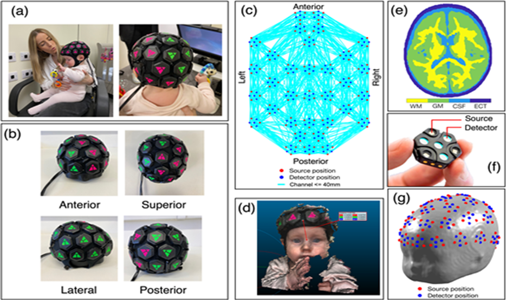
(a) 33-dock whole-head HD-DOT cap on a 7-month-old infant’s head. (b) Anterior, superior, lateral, and posterior views of the infant whole-head cap on a phantom. (c) Channel map for source-detector separations of 40 mm and below. (d) Anterior true-depth scan of an infant’s head, measuring the position of a point on the cap (intended to lie on the midline) relative to the nasion. (e) Axial slice of the tetrahedral volume mesh showing spatial distribution of white matter (WM), grey matter (GM), cerebrospinal fluid (CSF), and extra-cerebral tissue (ECT). (f) LUMO tile, consisting of four photodiode detectors and three dual-wavelength sources. Source-detector distances within each tile are 10 mm and 20 mm. (g) Source and detector positions registered to the infant scalp surface, for example, individual. (Collins-Jones et al. 2024)
Neuroscientists at the Centre for Brain and Cognitive Development (CBCD) at Birkbeck, University of London, were determined to use HD-DOT to understand the infant brain better. The team developed a whole-head infant HD-DOT system, which allowed them to measure brain activity across the entire surface of the infant’s scalp.
“Previous studies using HD-DOT had only been able to sample specific areas of the cortex,” researchers explained. “Our goal was to develop a system that could provide a more comprehensive picture of brain activity in infants.”
The team recruited 24 healthy, term-born infants aged between 5 and 7 months. The infants were placed on their parent’s lap and presented with audiovisual stimuli, including videos of actors performing nursery rhymes and moving mechanical toys. As the infants watched the videos, the HD-DOT system measured changes in blood flow and oxygenation in their brains.
The results were groundbreaking. The researchers identified specific brain regions that were activated in response to different types of stimuli. For example, they found that the brain’s temporal lobes were particularly active when infants were watching videos of people. In contrast, the occipital lobes were activated when they were watching videos of objects.
“We were surprised by the level of detail we could obtain from the HD-DOT data,” researchers said. “We could see subtle differences in brain activity that would have been difficult to detect using other methods.”
The researchers also found that the infants’ brains showed signs of specialisation at this early age. Different brain regions were activated in response to various stimuli, suggesting that the brain was beginning to develop specialised circuits for processing different kinds of information. By providing a more comprehensive picture of brain activity, this technique can help researchers to identify early signs of developmental disorders and to develop targeted interventions.
“We are just beginning to scratch the surface of what is possible with HD-DOT,” Scientist said. We are excited to see what we can learn about the infant brain in the coming years.”
As the researchers continued to analyse the data, they noticed something unusual. Some infants observed an inverted response in the sensorimotor regions of the brain. This meant these regions were becoming less active rather than more active during the task. The researchers hypothesised that the inverted response might be related to the infants’ developing motor skills. As the infants learned to control their movements, they may have been suppressing activity in the sensorimotor regions to avoid interfering with their voluntary actions.
To test this hypothesis, a follow-up study was conducted. They recruited new infants and presented them with the same audiovisual stimuli. However, this time, they also asked the infants to perform a simple motor task, such as reaching for a toy.
The results confirmed their hypothesis. When the infants were performing the motor task, they showed a reduced response in the sensorimotor regions of the brain. This suggested that the inverted response they had observed in the previous study was indeed related to the infants’ developing motor skills. This was believed to be a new aspect of infant brain development that had not been previously described. Their work highlighted the importance of studying the infant brain holistically, considering not only cognitive functions but also motor skills and other aspects of development.
As the researchers continued to explore HD-DOT’s potential, they began to consider how this technique could be used to study the development of atypical brains. They hypothesised that HD-DOT might be able to identify early signs of developmental disorders, such as autism spectrum disorder (ASD) or attention deficit hyperactivity disorder (ADHD).
To test this subsequent hypothesis, the researchers recruited a group of infants with ASD and compared their brain activity to that of typically developing infants. They found that infants with ASD showed distinct patterns of brain activity, suggesting that HD-DOT could be a valuable tool for early diagnosis and intervention, potentially helping to improve the lives of children with developmental disorders in the near future.
The findings from their study point to HD-DOT’s potential to revolutionise our understanding of the infant brain, unlocking the mysteries of early brain development, as well as aid in the development of new and innovative ways to support the healthy development of all children.
Conclusion
Whole-head HD-DOT offers a promising new tool for studying infant brain development. By providing a comprehensive picture of brain activity, this technique can help researchers to understand the complexities of the developing brain and to identify early signs of developmental disorders. As HD-DOT technology advances, we can expect to see even greater insights into the mysteries of the infant mind.
Disclaimer: The content provided on this platform is intended for informational purposes only and does not replace professional medical advice, diagnosis, or treatment. Some materials may originate from third-party sources, and the views expressed are solely those of the respective authors or entities, not Docquity. Docquity does not warrant the accuracy, reliability, or completeness of the content and disclaims any responsibility for it. Users are advised to independently verify all information before acting upon it.
Reference
Collins-Jones LH, Gossé LK, Blanco B, et al. Whole-head high-density diffuse optical tomography to map infant audiovisual responses to social and non-social stimuli. Imaging Neuroscience 2024;2:1–19. doi: 10.1162/imag_a_00244
About Docquity
If you need more confidence and insights to boost careers in healthcare, expanding the network to other healthcare professionals to practice peer-to-peer learning might be the answer. One way to do it is by joining a social platform for healthcare professionals, such as Docquity.
Docquity is an AI-based state-of-the-art private & secure continual learning network of verified doctors, bringing you real-time knowledge from thousands of doctors worldwide. Today, Docquity has over 400,000 doctors spread across six countries in Asia.
Meet experts and trusted peers across Asia where you can safely discuss clinical cases, get up-to-date insights from webinars and research journals, and earn CME/CPD credits through certified courses from Docquity Academy. All with the ease of a mobile app available on Android & iOS platforms!