In an extremely rare case, surgeons extracted a fetal mass from the brain of a one-year-old. The fetus in fetu discovery followed the child’s presentation of delayed motor skills, increased head size, and cerebral fluid accumulation.
The most recent case was disclosed in the American Academy of Neurology’s journal Neurology on December 12, 2022. The case revealed the presence of a ‘malformed monochorionic diamniotic twin’ within the child’s cranial cavity. These twins, originating from a single fertilized egg, share a placenta but possess separate amniotic sacs during gestation, indicating identical genetic makeup.
Fetus in Fetu: Medical Findings and Theoretical Explanations
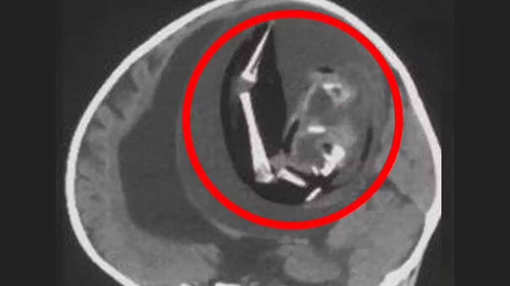
Doctors noted that the fetus had developed upper limbs, bones, and fingernails, suggesting ongoing growth within its sibling’s womb over several months. Measuring approximately four inches in length, the fetus remained undetected until the parents sought medical scans for their daughter due to her enlarged head and motor skill difficulties. The condition, termed “fetus in fetu” or occasionally “parasitic twin,” involves one fetus being enclosed by another, as per the reports. Typically, the absorbed twin ceases growth while the other continues to develop.

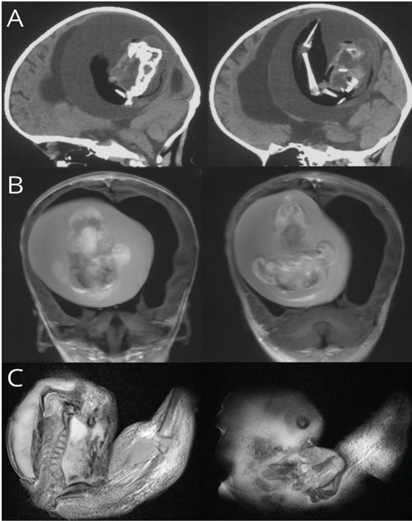
The above shows a scan of the infant girl’s skull with the fetus pictured inside.
Only around 200 cases have been documented, of which 18 occurred inside the skull.

Doctors said the fetus had developed upper limbs, bones, and even fingernails, meaning it likely continued growing for months while inside its sibling in the womb.
This rare phenomenon manifests in approximately 1 in 500,000 live births. Typically, the malformed fetus presents as a mass within the host fetus’s abdomen, nestled behind the abdominal wall tissues. However, in this instance, the mass was situated in the host fetus’s cranial cavity, likely originating very early in development during the blastocyst stage of the fertilized egg.
This condition arises from the incomplete separation of identical twins from a single egg division. The precise mechanism remains unknown to medical experts.
One theory proposes that the healthy twin establishes a connection with the mother’s placenta while the other twin attaches to the blood vessels of its sibling. Consequently, as the larger twin grows, the smaller one becomes absorbed into their abdomen. Alternatively, some scientists suggest it may occur due to delayed cell division.
Treatment and Long-Term Concerns
Despite being nonviable, the absorbed fetus might continue to develop for weeks or even months within its sibling, forming organs, bones, and limbs. The unnamed girl was admitted to the hospital due to motor skill issues, possibly affecting her ability to walk or sit.

While doctors didn’t provide specific details, her condition included the presence of her unborn sibling pressing against her brain, as revealed by CT scans. Additionally, she exhibited hydrocephalus, characterized by fluid accumulation deep within the brain, leading to symptoms like an enlarged head, excessive drowsiness, and seizures. Medical professionals noted that the survival of the fetus continued for a year post-birth because it shared a blood supply with its sibling. The potential for long-term damage to the surviving twin remained uncertain.
Dr. Zongze Li, a neurologist at Huashan Hospital, Fudan University, involved in the girl’s treatment, explained, “The intracranial fetus-in-fetu is believed to originate from unseparated blastocysts. The conjoined portions evolve into the host fetus’s forebrain and enclose the other embryo during neural plate folding.”
Historical Cases and Broader Implications
In 2017, medical professionals in Thailand discovered three siblings within the cranial cavity of an unborn girl. These siblings exhibited “multiple well-developed organs,” encompassing nervous, digestive, and respiratory systems. They were linked to their host sibling through a single artery and vein, which doctors identified as the umbilical cord.
Another incident from 2015, also in China, involved doctors discovering an unborn fetus within the scrotal sac of its male twin. Upon the 20-day-old infant’s birth, he was brought to the hospital due to swelling in his scrotum. Scans uncovered a “well-defined… mass” within the scrotum, containing bones and buds that doctors indicated would develop into limbs. Through surgery, the fetus was successfully removed, and the twin was discharged five days post-surgery, having fully recovered.
References
- In Extremely Rare Case, Doctors Remove Fetus from Brain of 1-Year-Old [Internet]. Accessed on June 10, 2024. Available at: https://reachmd.com/news/in-extremely-rare-case-doctors-remove-fetus-from-brain-of-1-year-old/2450453/
- Fetus Removed From Brain of 1-Year-Old Girl [Internet]. Accessed on June 10, 2024. Available at: https://www.medpagetoday.com/neurology/generalneurology/103475
About Docquity
If you need more confidence and insights to boost careers in healthcare, expanding the network to other healthcare professionals to practice peer-to-peer learning might be the answer. One way to do it is by joining a social platform for healthcare professionals, such as Docquity.
Docquity is an AI-based state-of-the-art private & secure continual learning network of verified doctors, bringing you real-time knowledge from thousands of doctors worldwide. Today, Docquity has over 400,000 doctors spread across six countries in Asia. Meet experts and trusted peers across Asia where you can safely discuss clinical cases, get up-to-date insights from webinars and research journals, and earn CME/CPD credits through certified courses from Docquity Academy. All with the ease of a mobile app available on Android & iOS platforms!


