Introduction
Conjoined twins, an extraordinary medical phenomenon, have long fascinated both the medical community and the general public. Among the array of conjoined twin variations, heteropagus conjoined twins stand as an even rarer occurrence. Distinguished by their asymmetry, heteropagus twins comprise one nearly normal twin (autosite) and another incompletely formed twin physically attached to the autosite (parasite). A subset of this anomaly, known as Epigastric Heteropagus Conjoined Twins (EHTs), in which the parasitic twin is connected to the epigastrium of the autosite, is an exceptionally uncommon occurrence.1
Scarcity of Knowledge
The scarcity of EHT cases has translated into limited scientific literature, contributing to a deficiency of understanding among medical practitioners regarding the optimal management of such cases. Compounding the issue, EHTs often present complex medical scenarios, contrasting typical conjoined twins. Consequently, the etiology, presentation, and therapeutic outcomes of EHTs remain subjects of ongoing debate. This article sheds light on this perplexing condition by presenting two cases of EHTs observed within a brief time frame, offering insights into their manifestation, surgical treatment, and genetic underpinnings.
Case Studies2
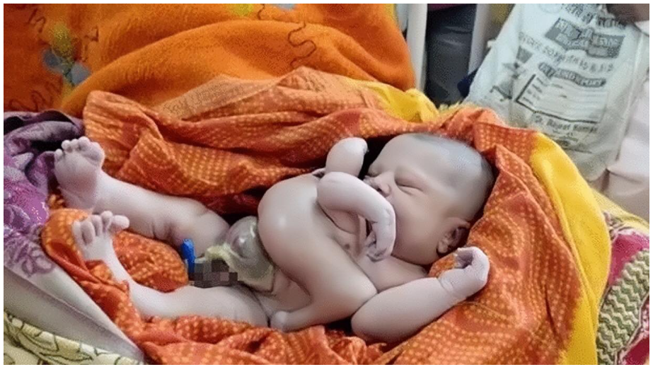
Case 1: A 1-day-old male was born via full-term normal delivery, presenting with a parasitic twin attached to the epigastrium of the autosite. The parasitic twin exhibited various anatomical features, including upper and lower limbs, a well-developed scrotum and penis, and urinary function. Surgical separation took place during the third month after birth. The parasitic twin displayed cartilage connections to the autosite’s sternum and shared an intricate liver connection. This case demonstrated the intricate vascular, skeletal, and anatomical connections that typify EHTs, alongside the absence of skeletal muscle in the parasitic twin’s limbs. Post-separation, the autosite progressed without complications over 52 months (Figure 1).

Figure 1: (A) The parasitic twin in case 1 had a fully developed pelvis, two pairs of limbs, and a well-developed penis, which produced urine discharge. (B) The livers of the parasite and autosite were physically attached in case 1. (C) The parasitic twin in case 1 contained a pair of well-developed kidneys. (D) The autosite in case 1 during the twelfth month after the operation to remove the parasitic twin.

Figure 2: (A) Large hernia of the abdominal wall (H), omphalocele (O) and limbs (L) of the parasitic twin in case 2. (B) View of the hernia in case 2. The parasitic twin was excised, and the omphalocele was repaired. (C) The pelvis of the parasitic twin in case 2 contained one small kidney and a cyst. (D) The hernia in case 2 was repaired using VYPRO II mesh. (E) Condition of the repaired ventral hernia in case 2 at 14 days after surgery. (F) The autosite in case 2 during the twenty-third month after the operation to remove the parasitic twin.

Figure 3: Case 2. Results of the DNA analysis of the twins in case 2. (A) Skin from the autosite. (B) Skin from the parasite. (C) Hair from the autosite. (D) Kidney from the parasite.
Case 2: Another male patient born on the same day exhibited a distinct manifestation of EHT, encompassing a parasitic twin with immobile lower limbs and an infected omphalocele attached to the epigastrium of the autosite. The autosite featured an infected omphalocele and a large ventral hernia. The omphalocele was repaired shortly after birth, followed by successfully removing the parasitic twin. This case showcased the challenge of managing concurrent hernia and separation surgeries while avoiding complications. Notably, the DNA analysis of this case revealed a monozygotic origin between the autosite and the parasitic twin, contributing to the evolving genetic understanding of EHTs (Figure 2).
Incidence and Origins
Conjoined twins, though rare, have varying reported incidence rates across studies. The global range is 1 in 50,000 to 200,000 births, with heteropagus twins constituting 10% of this subset.3 The complexity of EHTs and their infrequency results in limited statistical data on these cases. The etiology of conjoined twins remains a mystery, with speculation surrounding spontaneous generation from an error in blastogenesis during the early stages of human development. Emerging theories, such as a “fusion theory,” present alternate possibilities for the origin of heteropagus twins. As seen in Case 2, genetic analysis advances our understanding of EHT origins.
Gender Bias, Skeletal Atrophy, and Anatomical Associations
EHTs display a gender bias, with males constituting approximately 78% of cases. Skeletal muscle atrophy, leading to the absence of proper innervation, contributes to the limitations of movement observed in the parasitic twin’s limbs. Intriguingly, both cases highlighted cartilage connections to the autosite’s sternum, suggesting a potential origin from active centres of cell proliferation like the sternum, cutis, and pelvis. Moreover, omphaloceles appeared as common accompanying anomalies in EHTs, with underlying mechanisms positing interference with abdominal wall closure due to connecting cartilage bridges.
Surgical Complexity and Optimistic Prognosis
The rarity of EHTs underscores the complexity of their management. In the presented cases, surgical separation succeeded, reflecting the critical role of early diagnosis, meticulous prenatal care, and well-planned surgical strategies. Notably, the large hernia case highlighted the challenges of simultaneous hernia repair and separation. A calculated delay in hernia repair was implemented to avoid potential complications. Encouragingly, both autosites showed normal development post-surgery, reinforcing the potential for positive outcomes in EHT cases.
Conclusion
Epigastric Heteropagus Conjoined Twins remain a captivating enigma within the realm of medical anomalies. Through analyzing two unique cases, this article delved into the complexities surrounding their aetiology, presentation, and management. The rarity of EHTs and their diverse manifestations underscore the need for continued research, fostering a deeper understanding of these exceptional occurrences and advancing the potential for effective diagnosis and treatment.
References
- Bhansali M, Sharma DB, Raina VK. Epigastric heteropagus twins: 3 cases reports with review of literature. Journal of Pediatric Surgery. 2005;40(7):1204–8.
- Tongsin A, Niramis R, Rattanasuwan T. Epigastric hateropagus twins: a report of four cases. J Med Assoc Thai. 2003;86(Suppl 3):S605–9.
- Kallen B, Rybo G. Conjoined twins in Sweden. Acta Obstet Gynecol Stand. 1978;57(3):257–9.